Lower Back Bones Diagram : The bones of the lower back - Stock Image - F001/3881 ... - Without hands on experience hip bone is very tough to understand but you made it very simple thank you mam most underrated youtube channel with good videos.
Lower Back Bones Diagram : The bones of the lower back - Stock Image - F001/3881 ... - Without hands on experience hip bone is very tough to understand but you made it very simple thank you mam most underrated youtube channel with good videos.. Lower jaw (mandible) collar bone. These bones work together to provide flexibility to the trunk, support the muscles of the trunk, and protect the spinal cord and spinal nerves of the back. Anatomy the american center for spine and neurosurgery. Fitness and low back pain physiopedia. The spinal cord is to learn more about the anatomy of the spine, watch this video.
Find the perfect bone diagram stock illustrations from getty images. Diagram of back bones wiring. This framework consists of many individual bones and cartilages. Click here to read about mesothelioma and its differential diagnosis and mesothelioma treatments. This bone is shaped like a triangle that fits between the two halves of the pelvis connecting the spine to the lower half of the body.
Pelvis | Pelvis anatomy, Hip anatomy, Medical anatomy from i.pinimg.com The mandible sits beneath the maxilla. The bones of the pelvis and lower back work together to support the body's weight, anchor the abdominal and hip muscles, and protect. Fitness and low back pain physiopedia. Lower back pain back bones lumbar stock illustration 679275577. Bone science human diagram anchor chart human body health back skeleton. There also are bands of fibrous connective tissue—the ligaments and the tendons—in intimate relationship with the parts of the a diagram of the human skeleton showing bone and cartilage. Back bones structure bone structure lower back humananatomybody. 3,406 bone diagram stock illustrations and clipart.
This bone is shaped like a triangle that fits between the two halves of the pelvis connecting the spine to the lower half of the body.
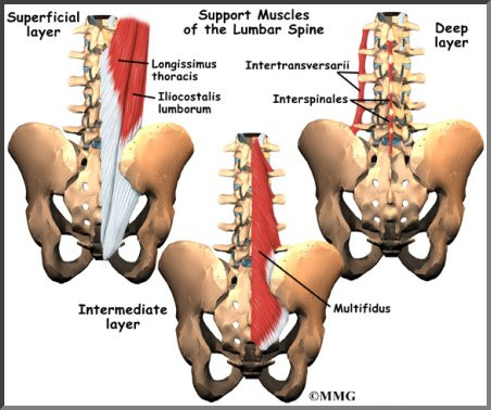
Spinal anatomy spine treatment boulder neurosurgical spine. Lower back muscle diagram anatomy. Understanding lower back anatomy is key to understanding the root of lower back and hip pain. In this image, you will find an occipital bone, sternocleidomastoid, trapezius, deltoid in muscles of the lower back diagram. Diagram of back bones wiring. Nice lower back anatomy diagram ornament internal organs diagram. Cheek bone (zygoma) upper jaw (maxilla). Start studying skeletal diagram, bone information. You can strain these muscles by stretching them too. Click here to read about mesothelioma and its differential diagnosis and mesothelioma treatments. A series of muscles and ligaments in your back hold the bones of your spinal column in place. The pain usually results from problems with the musculoskeletal system—most notably the spine, including the bones of the spine (back bones, or vertebrae), disks, and the muscles and ligaments that support it. Bone science human diagram anchor chart human body health back skeleton.
Low back pain and neck pain are among the most common reasons for health care visits. The bones of the pelvis and lower back work together to support the body's weight, anchor the abdominal and hip muscles, and protect. Your lower back contains 5 vertebral bones stacked above each other with intervertebral discs in between. Human back muscles and bones, human backbone structure. The spinal cord is to learn more about the anatomy of the spine, watch this video.
vertebrae | ... vertebrae, 5 lumbar vertebrae, 1 sacrum (5 ... from i.pinimg.com The lumbar and sacrum region make up the bone of the lower back anatomy. Free download abdomen,spleen,liver anatomy and physiology diagrams. Cheek bone (zygoma) upper jaw (maxilla). Low back pain and neck pain are among the most common reasons for health care visits. You can strain these muscles by stretching them too. Spinal anatomy spine treatment boulder neurosurgical spine. For example, the long bones of the lower arm and the leg both have attached interosseous membranes. In this image, you will find an occipital bone, sternocleidomastoid, trapezius, deltoid in muscles of the lower back diagram.
The bones of the pelvis and lower back work together to support the body's weight, anchor the abdominal and hip muscles, and protect.
Fitness and low back pain physiopedia. 3,406 bone diagram stock illustrations and clipart. In this image, you will find an occipital bone, sternocleidomastoid, trapezius, deltoid in muscles of the lower back diagram. Your lower back (lumbar spine) is the anatomic region between your lowest rib and the upper part of the buttock.1 your spine in this region has a natural inward curve. Cheek bone (zygoma) upper jaw (maxilla). Low back pain is a fact of life. Lower back muscle diagram anatomy. Lower back pain back bones lumbar stock illustration 679275577. The spinal cord is to learn more about the anatomy of the spine, watch this video. Lower jaw (mandible) collar bone. I'm going through all your bone tutorials. Understanding lower back anatomy is key to understanding the root of lower back and hip pain. Free download abdomen,spleen,liver anatomy and physiology diagrams.
So what is low back strain? For example, the long bones of the lower arm and the leg both have attached interosseous membranes. I'm going through all your bone tutorials. It forms the lower jaw and holds the lower teeth in place. 3,406 bone diagram stock illustrations and clipart.
Lumbar Spine Anatomy | eOrthopod.com from eorthopod.com So what is low back strain? As you can see, there are also have a spine of scapula deltoid, triceps brachii, latissimus dorsi. The mandible sits beneath the maxilla. This framework consists of many individual bones and cartilages. Lower back bones diagram : Cheek bone (zygoma) upper jaw (maxilla). Click here to read about mesothelioma and its differential diagnosis and mesothelioma treatments. In this image, you will find an occipital bone, sternocleidomastoid, trapezius, deltoid in muscles of the lower back diagram.
Click here to read about mesothelioma and its differential diagnosis and mesothelioma treatments.
The mandible sits beneath the maxilla. Find the perfect bone diagram stock illustrations from getty images. Learn vocabulary, terms and more with flashcards, games and other study tools. While many conditions can lead to lbp, inadequate core strength is a common causal factor. Would you like to tell us about a lower price? Brief content visible, double tap to read full content. Just about everybody will suffer from it sooner or later. Spinal anatomy spine treatment boulder neurosurgical spine. Lower jaw (mandible) collar bone. This picture also contains humerus, olecranon process of ulna, deep to tendon and. Anatomy the american center for spine and neurosurgery. Low back pain and neck pain are among the most common reasons for health care visits. Cheek bone (zygoma) upper jaw (maxilla).
The bones of the pelvis and lower back work together to support the body's weight, anchor the abdominal and hip muscles, and protect back bones diagram. Low back pain is a fact of life.


0 Komentar